With sleep apnea affecting breathing patterns and jaw alignment, your dentist can spot oral signs that indicate your risk-worn tooth surfaces, a scalloped tongue, dry mouth, inflamed gums, and an enlarged soft palate-and will assess your airway anatomy, occlusion, and bite to determine whether sleep-disordered breathing contributes to your oral health issues.
Understanding Sleep Apnea
Definition and Types
Sleep apnea causes repeated pauses in your breathing during sleep; obstructive (OSA) stems from upper airway collapse, central (CSA) from reduced respiratory drive, and complex or mixed forms combine both mechanisms, so your dental findings – like jaw position or palatal anatomy – help differentiate which pathway is involved.
| Type | Key features relevant to dental exam |
| Obstructive (OSA) | Loud snoring, retrognathia, large soft palate, daytime sleepiness; often responsive to oral appliance therapy |
| Central (CSA) | Minimal snoring, neurologic contributors, variable jaw findings; dental role is limited to symptom screening and coordination |
| Mixed/Complex | Combined collapse and central drive issues; requires multidisciplinary management and careful appliance selection |
| UARS/positional | Upper airway resistance with flow limitation or position-dependent events; dental posture and mandibular advancement can influence severity |
- Palatal scalloping, large tonsils, and a narrow maxillary arch often point toward airway obstruction.
- Bruxism and morning headache increase the index of suspicion in your dental exam.
- Any loud, witnessed apneas or frequent choking arousals you report should trigger screening and referral.
Prevalence and Risk Factors
Across adults, OSA prevalence varies by age, sex, and BMI; estimates range from about 10% in middle-aged populations to higher rates in older adults, with moderate-to-severe OSA affecting roughly 10% of middle-aged men and increasing markedly with obesity-if you have a BMI over 30 your likelihood rises substantially.
- Obesity: each 10% weight gain raises OSA risk markedly.
- Age and male sex: prevalence increases after 40 and is higher in men.
- Craniofacial anatomy: retrognathia, narrow maxilla, or large tonsils elevate risk.
- Lifestyle: alcohol use, sedatives, and smoking worsen airway collapsibility.
- Assume that a combination of factors (e.g., BMI >30 plus loud snoring) meaningfully increases pretest probability.
In dental practice audits and screening cohorts, you’ll often find a high rate of undiagnosed OSA among patients presenting with bruxism, morning dry mouth, or a scalloped tongue; screening tools (STOP-Bang, Epworth) applied in-chair can identify those with >50% probability, and targeted referrals reveal a substantial proportion who require CPAP or oral appliance therapy.
- Screen for loud habitual snoring, witnessed apneas, and daytime sleepiness during your exam.
- Measure neck circumference and assess craniofacial structure using photos or intraoral records when available.
- Note comorbid hypertension, atrial fibrillation, or insulin resistance that often co-occur with OSA.
- Document bruxism and tooth wear patterns that may signal nocturnal arousal events.
- Assume that identifying two or more objective risk markers warrants discussion of sleep testing and sleep-medicine referral.
The Impact of Sleep Apnea on Oral Health
Altered breathing and sleep fragmentation shift your oral environment: mouth breathing lowers saliva-studies report 30-50% of people with obstructive sleep apnea (OSA) experience dry mouth-while repetitive arousals increase bruxism and TMJ stress. You may notice accelerated enamel wear, rising caries rates, and denture instability; your dentist can screen for these signs and offer therapies such as oral appliance therapy, which often reduces apnea events by roughly 50%. See What Your Dentist Can Do For Sleep Apnea and Oral Health.
Common Oral Health Issues
You frequently present with increased cavities from reduced saliva, enamel abrasion from nightly grinding, and heightened tooth sensitivity; clinical data show bruxism and enamel wear are markedly higher in OSA patients. Dentures and crowns loosen sooner when tissues are persistently dry, and CPAP-related mouth leaks can exacerbate mucosal irritation and halitosis, so your dentist will inspect restorations and soft tissues at follow-up.
Effects on Gum Health
You face an elevated risk of periodontal disease: research links OSA with roughly a twofold increase in periodontitis prevalence. Intermittent hypoxia drives systemic inflammation, producing higher IL‑6 and CRP levels that manifest orally as deeper pockets, more bleeding on probing, and slower healing after periodontal therapy.
More detailed studies show moderate-to-severe OSA is associated with a 1.5-2.0-fold greater odds of advanced periodontitis after adjusting for age, smoking and BMI. Your dentist will measure pocket depths, attachment loss and bleeding on probing, and may coordinate OSA treatment because reducing nocturnal hypoxia can lower inflammatory markers and improve periodontal treatment outcomes.
Signs Dentists Look for in Patients
When assessing your mouth and airway, dentists watch for visible clues, patient reports, and bite patterns that suggest sleep-disordered breathing; for more on what clinicians note first see Oral Health & Sleep Apnea: What Your Dentist Might See …-these findings guide screening, documentation, and referrals for sleep testing.
Physical Indicators
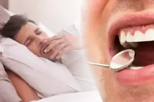
Your dentist inspects for a high-arched or narrow palate, retrognathia or small jaw, enlarged tonsils or uvula, scalloped tongue, and excessive tooth wear; they also note crowding, a deviated septum visible on oral exam, and soft-tissue hypertrophy that can reduce airway space during sleep.
Symptoms Related to Sleep Apnea
You may report loud snoring, witnessed pauses in breathing, frequent nighttime awakenings, dry mouth on waking, or excessive daytime sleepiness-symptoms that, when combined with oral findings, raise suspicion for obstructive sleep apnea (OSA).
To clarify risk, your dentist often uses brief screens like the 8-item STOP-Bang or the Epworth Sleepiness Scale (scores >10 suggest significant sleepiness), asks about partner observations, and documents symptom frequency to decide on referral for home sleep apnea testing or in-lab polysomnography.
The Role of Dentists in Diagnosis
When you sit for a dental exam, your dentist assesses bite, jaw position, tongue scalloping, tonsil size and airway space-alongside BMI and neck circumference-to flag sleep apnea risk; they also note bruxism and REM-related wear patterns. You should expect documentation that can be used by a sleep specialist: apnea-hypopnea index (AHI) thresholds are 5-15 (mild), 15-30 (moderate), and >30 (severe), which helps determine whether you need referral for testing or an oral appliance trial.
Screening Techniques
You will often complete validated tools such as STOP-Bang (score ≥3 suggests high risk) or the Epworth Sleepiness Scale; dentists perform Mallampati staging, Friedman tongue position, and intraoral photography. Portable pulse oximetry or home sleep apnea tests (HSAT) may be ordered or coordinated with a physician when you present with daytime sleepiness, witnessed apneas, or multiple risk factors like BMI >30 and neck circumference above 17 inches in men.
Collaboration with Sleep Specialists
You rely on coordinated care: after a positive screen your dentist typically refers you for polysomnography or HSAT, then fabricates a mandibular advancement device (MAD) only with a physician diagnosis and prescription. Follow-up is shared-sleep physicians track AHI changes while your dentist manages fit, titration and dental side effects-because effective therapy, especially for moderate OSA, requires both diagnostic confirmation and device optimization.
In practice, your dentist will exchange reports and device progression notes with the sleep specialist, including baseline AHI, symptom logs, and appliance advancement records (commonly adjusted in 0.5-1 mm steps). You should expect follow-up at 1-3 months for symptom review, repeat HSAT or oximetry to document AHI reduction (MADs often lower AHI by about 50%), and annual dental exams to monitor occlusion and tooth movement while the sleep team assesses long-term respiratory outcomes.
Treatment Options for Sleep Apnea
You can choose from CPAP, oral appliances, positional therapy, or surgery depending on severity and anatomy; CPAP often lowers the apnea-hypopnea index (AHI) by more than 70% with good use, while mandibular advancement devices cut AHI by roughly 30-60% in mild-moderate cases. Consult your dentist and sleep specialist for coordinated care and see Spotting Dental Signs of Sleep Apnea in Philadelphia …
Oral Appliances
You’ll usually get a custom mandibular advancement device from a dentist trained in sleep medicine; it advances your lower jaw 5-10 mm to open the airway, and studies report symptom improvement in about 50-70% of mild-moderate cases. Expect follow-ups for bite changes, TMJ monitoring, and periodic adjustments-overnight sleep studies or home testing can verify effectiveness.
CPAP Therapy
You should view CPAP as the gold-standard for moderate to severe obstructive sleep apnea: continuous positive airway pressure splints the airway open, and adherence of ≥4 hours per night on most nights is linked to the largest health benefits, including reduced daytime sleepiness and lower cardiovascular risk.
Mask choice matters for you-nasal, nasal pillows, or full-face masks fit different breathing patterns and facial anatomy; pressure can be fixed or auto-adjusting (APAP), with typical therapeutic ranges from 4-20 cm H2O. If you struggle with leaks, dry mouth, or aerophagia, heated humidification, a ramp feature, or mask refitting often helps. Medicare defines adherence as ≥4 hours on 70% of nights over 30 days, so early troubleshooting with your provider and coordinated care with your dentist (if you use a simultaneous oral appliance) improves long-term success.
Preventive Measures for Oral Health
Proactive daily care and targeted follow-up stop small problems from becoming big ones: brush twice daily for two minutes with fluoride toothpaste, floss once a day, clean your tongue, replace your toothbrush every three months, and hydrate to reduce nighttime dry mouth; if you use an oral appliance or CPAP, clean and inspect devices nightly and track symptoms so your dentist can intervene early.
Dental Hygiene Practices
You should brush for two minutes twice daily with a soft-bristle brush and fluoride toothpaste, floss or use interdental cleaners once a day, and use a tongue scraper to cut bacterial load; for dry mouth consider saliva substitutes and alcohol-free mouthwash, and clean any oral appliance nightly with manufacturer-approved methods to prevent biofilm buildup and halitosis.
Regular Check-Ups
Plan dental visits at least every six months, and if you wear a mandibular advancement device schedule a 4-6 week follow-up after fitting and then every 3-6 months during titration; these visits let your dentist catch periodontal disease, tooth wear, appliance fit issues, and early bite changes before they worsen.
During follow-ups your dentist will chart occlusion, measure pocket depths, check for bruxism and TMJ pain, assess airway indicators like tongue scalloping and tonsil size, update radiographs as needed, and use screening tools (for example STOP-Bang-style questions) to decide if a sleep study or ENT referral is warranted; detailed baseline records and periodic photos help quantify appliance-related tooth movement and guide timely adjustments.
To wrap up
Taking this into account, when assessing how sleep apnea affects your oral health your dentist will look for signs such as tooth wear from bruxism, enamel erosion, dry mouth, periodontal inflammation, a scalloped tongue, crowded or retrognathic anatomy, and evidence of mouth breathing; addressing these findings helps you reduce airway obstruction risks and coordinate treatment with sleep specialists.
FAQ
Q: How can dentists spot possible sleep apnea signs during a routine exam?
A: Dentists look for patterns such as severe enamel wear from bruxism, scalloped tongue edges, enlarged tonsils or a narrow oropharynx, and a high-arched or narrow palate. They check for intraoral dryness, cracked or chapped lips, and evidence of long-term mouth breathing. Facial profile cues like retrognathia (receded lower jaw) and reduced posterior airway space on imaging also raise suspicion.
Q: What oral health problems are commonly linked to sleep apnea?
A: Commonly associated problems include bruxism-related tooth wear, increased risk of periodontal disease, xerostomia (dry mouth) often from mouth breathing or CPAP use, and temporomandibular joint (TMJ) pain from oral appliance therapy or clenching. Chronic inflammation from intermittent hypoxia can worsen gum disease and impair healing after dental procedures.
Q: What patient history and screening tools do dentists use to evaluate sleep apnea risk?
A: Dentists gather sleep history (snoring, witnessed apneas, daytime sleepiness), medical history (hypertension, obesity), and lifestyle factors. Standard screening tools include the STOP-BANG questionnaire and the Epworth Sleepiness Scale; positive screens prompt referral for definitive testing. Neck circumference, BMI, and reports from bed partners are also factored into risk assessment.
Q: Which intraoral and craniofacial features most strongly suggest obstructive sleep apnea?
A: Key features include a small or retruded mandible, large tongue or tongue scalloping, tonsillar hypertrophy, a narrow/high-arched palate, crowded dental arches, and a low tongue posture. A Mallampati-style assessment of oropharyngeal visibility and reduced posterior airway space on cephalometric or CBCT imaging provide objective support for concern.
Q: How do dental oral appliances for sleep apnea work and what side effects should be monitored?
A: Mandibular advancement devices (MADs) advance the lower jaw to enlarge the airway and reduce collapsibility during sleep. Dentists must titrate advancement, monitor for jaw pain, bite changes, tooth movement, excessive salivation or dry mouth, and TMJ symptoms. Regular follow-ups and occlusal checks are needed to manage progressive occlusal changes and ensure continued efficacy.
Q: In what ways does untreated sleep apnea affect periodontal health and tooth survival?
A: Intermittent nocturnal hypoxia and sleep fragmentation increase systemic and local inflammation, which can accelerate periodontal attachment loss and bone resorption. Mouth breathing and xerostomia reduce salivary protection, raising caries risk and altering oral microbiota. These factors together can lead to faster progression of gum disease and higher risk of tooth loss without appropriate management.
Q: When should a dentist refer a patient for medical sleep evaluation and how do they coordinate care?
A: Dentists should refer patients who have positive screening results, significant craniofacial risk factors, loud habitual snoring, witnessed apneas, or unexplained daytime sleepiness. Coordination typically involves recommending a sleep physician for polysomnography or home sleep apnea testing, sharing dental findings and imaging, and discussing possible oral appliance therapy as part of a combined treatment plan. Ongoing communication ensures appliance fitting, medical treatment (e.g., CPAP), or surgical options are integrated and outcomes are monitored.